
A New Path for UBE minimally invasive Technology
Author: TL-WC
Release time: 2026-01-16 05:10:44
View number: 1464
New approach of UBE minimally invasive technology: Precise treatment of extremely lateral lumbar intervertebral disc protrusion through paraspinal approach
In the field of minimally invasive spinal surgery,extraforaminal lumbar intervertebral disc protrusionDue to its special anatomical location - on the outside of the intervertebral foramen and adjacent to the outlet nerve root - it often causes severe radiating lower extremity pain and has a poor response to conservative treatment. Although traditional open surgery is effective, it requires extensive muscle dissection and partial resection of the superior articular processes, which may affect the long-term stability of the spine.
In recentUnilateral dual-channel endoscopic technique(Unilateral Biportal Endoscopy, UBE) provides a better minimally invasive solution for this type of lesion with its advantages of high-definition vision, flexible operation and tissue protection. Among them,The approach is via the paravertebral muscle spaceThe paraspinal approach is gradually becoming a new option for advanced surgeons due to its high degree of preservation of the articular process structure.
I. Why Choose the paravertebral Approach?
The extremely lateral type protrusion mostly occursL4-L5 and above segmentsThe lesion is located on the lateral side of the pedicle and in front of the transverse process. If the classic interlaminal approach is adopted, excessive lateral traction or grinding of the superior articular process is required, which can easily damage the small joint capsule and increase the risk of postoperative instability.
whileParavertebral approachEntering from the interstitial space between the multifidus and longus muscles, one can reach the transverse process - isthmus areaBypass the laminaDirectly expose the lateral area of the intervertebral foramen to achieve decompression with almost no damage to the superior articular processes.
Ii. Key Anatomical Localization: The "Safety Triangle" Principle
The core of successfully implementing this approach lies in identifying the "working triangle" composed of three bony landmarks:
The "C-shaped area" formed by the three is the safety window for surgical operations. Limited bony fenestration in this area can not only fully expose the nerve roots but also preserve the integrity of the articular processes to the greatest extent.
Iii. Brief Description of the Surgical Procedure
Note: Due to the iliac crest obstruction in the L5-S1 segment, the working channel can be slightly moved inward to optimize the operating Angle.
Iv. Summary of Clinical Advantages
V. Suggestions for Applicable Scenarios
This technology is applicable to:
⚠️ Note: The operator should have a solid foundation in spinal anatomy and UBE operation experience. It is recommended to carry out the operation after standardized training.
References direction(Not directly quoted, only for background support) :
In recent years, many international studies (such as EFORT Open Reviews and Global Spine Journal) have pointed out that the parapvertebral approach of UBE has shown good safety and efficacy in the treatment of high extreme lateral intervertebral disc protrusion, especially suitable for young patients who pay attention to facet joint protection.
In recent years, many international studies (such as EFORT Open Reviews and Global Spine Journal) have pointed out that the parapvertebral approach of UBE has shown good safety and efficacy in the treatment of high extreme lateral intervertebral disc protrusion, especially suitable for young patients who pay attention to facet joint protection.

Related Products
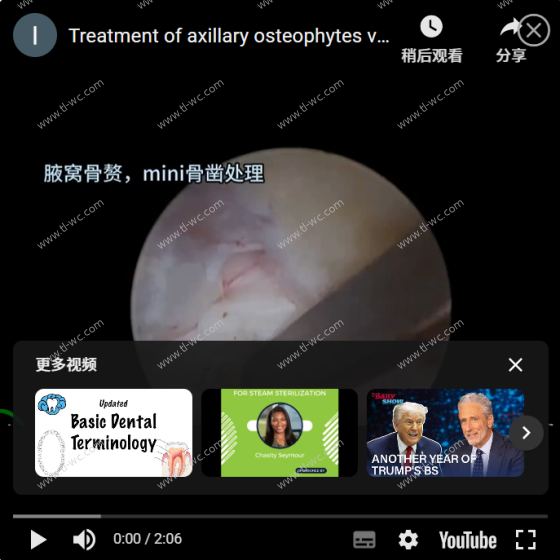
UBE surgery: Cervical corner approach keyhole, axillary osteophyte treatment, mini Angle bone knife and small grinding head
$0.00
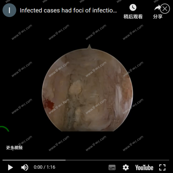
UBE surgery case: An infection case, with infection foci on both the upper and lower endplates of the L4 vertebra. Surgical operation video
$0.00
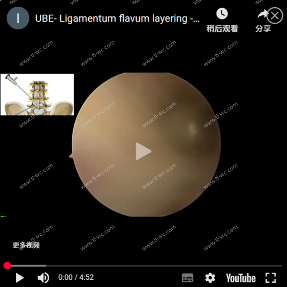
UBE- Ligamentum flavum layering -Corner area enters the spinal canal to operate AYDEFY
$0.00
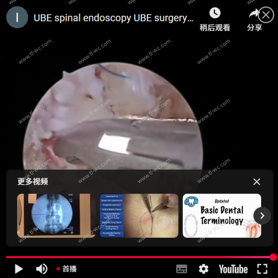
UBE spinal endoscopy UBE surgery annulus fibrosus suture video
$0.00